The S-protein receptor binding domain (RBD) of SARS-CoV-2 has been the subject of a thorough characterization and its importance in virus infection is well known. The potential uses of RBD of S-protein as a vaccine and as an antigen for the specific and sensitive detection of SARS-CoV-2 antibodies are herein reviewed.
The cell-free reaction is basically transcription and translation occurring in a test tube outside of the cell using the cellular machinery from inside. Hence, the technique relies on cellular extracts to provide the apparatus required for “cell free” protein expression. This cellular lysate is cleared out of DNA and mRNA, then supplemented with an energy supply and additives such as RNA polymerase to facilitate synthesis of proteins. The origin of the extract can be a variety of sources including prokaryotic, archaeal, fungal, plant, insect, mammalian or bacterial cells.
SARS-CoV-2
Severe acute respiratory syndrome coronavirus 2 (SARS-CoV-2) belongs to the β-coronavirus genus and shows close association with some bat coronaviruses and with SARS-CoV [1,2] being, though, more easily transmitted between humans than the SARS-CoV. This high transmission feature justifies its rapid spread throughout the planet and the setting up of the current COVID-19 pandemic [3].
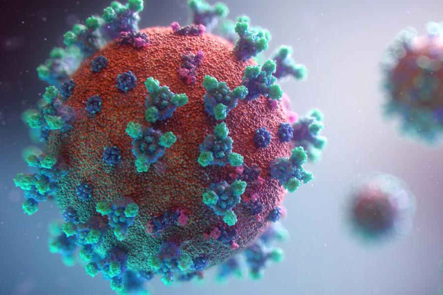
The SARS-CoV-2 virion comprises a nucleocapsid core together with an envelope that contains three membrane proteins named spike (S), envelope (E) and membrane (M) proteins [4]. S-protein plays a pivotal role in virus infection, being involved in viral attachment, fusion and entry in host cells, by interacting with a host receptor (angiotensin converting enzyme 2 – ACE2). The critical function of S-protein has led to consider it a potential target for the development of vaccines, entry inhibitors and antibodies anti-SARS-CoV-2 [5,6].
Interaction between the virus and ACE2 is performed through the receptor binding domain (RBD) located in the S1 subunit of S-protein, followed by fusion of viral and host membranes by the S2 subunit [7]. To fuse the viral and the host cell membranes, S-protein goes through a structural rearrangement [8,9], triggered when S1 unit binds to ACE2. This results in the shedding of the S1 unit and transition of the S2 unit to a stable conformation [10].
RBD in S-Protein : a flexible segment
The RBD in the S-protein, with a 193-residue length, is the most flexible segment in the CoV structure [11], and several RBD residues have shown to be essential to the interaction of S-protein with ACE2 [12,13]. In order to enable the connection with the host cell receptor, RBD undergoes hinge-like conformational movements that cause transient hiding or exposing of the receptor binding determinants. The conformations are known as “down” and “up” conformations, corresponding to an inaccessible receptor state and to an accessible receptor state, respectively [14]. This triggering mechanism is thought to be conserved among the coronaviruses [15,16].
It was found that S-protein from β-CoV has a common ancestor and originates from the outer region of the RBD, determining the potential of the virus for interspecies transmission [11]. RBD is responsible for the similar binding mechanism among the different coronaviruses, with the general conformation of S-protein being identical for SARS-CoV and SARS-CoV-2, with minor differences [14], and with RBD showing a 73% identity in protein sequence for SARS-CoV and SARS-CoV-2 [17].
RBD : a good target ?
Based on this similarity, several studies have been performed in order to evaluate the possibility to use anti-SARS-CoV monoclonal antibodies to neutralize SARS-CoV-2 RBD. It was found that a SARS-CoV-specific human monoclonal antibody named CR3022, isolated from a convalescent SARS patient, could bind to SARS-CoV-2 RBD in a potent way, although other SARS-CoV-specific antibodies did not react with the SARS-CoV-2 S-protein, which may be due to differences in RBD of both viruses [17]. The diverse behaviour of the tested antibodies may be due to the fact that CR3022 recognizes an epitope that does not overlap with the ACE2 binding site of SARS-CoV-2 RBD. The differences in RBD between SARS-CoV and SARS-CoV-2 do not impair the ability to bind to ACE2, but have a more pronounced effect on the cross-reactivity of neutralizing antibodies. Therefore, novel monoclonal antibodies specific for SARS-CoV-2 RBD are needed. Moreover, these results show that the RBD within S-protein is the critical target for neutralizing antibodies [18].
In another study [19], a recombinant SARS-CoV-2 RBD protein was shown to bind strongly to human and bat ACE2, and it also blocked the entry of SARS-CoV-2 and SARS-CoV into human ACE2-expressing cells in a dose-dependent manner, suggesting its potential use as a viral attachment inhibitor to prevent infection by both viruses. Furthermore, some cross-reactivity was observed for SARS-CoV RBD-specific antibodies towards SARS-CoV-2 RBD protein, inhibiting SARS-CoV-2 entry into human ACE2 cells. These results open the possibility to develop either a SARS-CoV or a SARS-CoV-2 RBD-based vaccine for SARS-CoV and SARS-CoV-2 infection.
RBD based vaccine
To assess the potential of a vaccine based on SARS-CoV-2 RBD, a study was carried out using a recombinant vaccine containing residues 319-545 of RBD within S-protein [20]. The results showed that the developed vaccine induced a potent antibody response in immunized mice, rabbits and non-human primates, in the first 7-14 days from injection of a single vaccine dose, in a dose-dependent manner. Sera of the immunized animals blocked the binding of RBD to ACE2 and neutralized the infection by a SARS-CoV-2 pseudovirus and live SARS-CoV-2 in vitro. Furthermore, the antigenicity of the recombinant vaccine was evaluated by testing seroreactivity in sera from 20 healthy donors and from 16 patients with COVID-19. All the 16 samples from COVID-19 patients showed significantly elevated levels of IgG and IgM against the recombinant RBD vaccine, compared to the results from healthy individuals. The recombinant vaccine showed a viral neutralization effect higher than other vaccines comprising the S1 or the S2 subunits, demonstrating the importance of including RBD in the design of SARS-CoV-2 vaccines. Toxicity evaluation was performed in the non-human primates and no adverse effects were observed.
RBD antigen for SARS-CoV2 diagnosis
In addition, to demonstrate the usefulness of RBD from S-protein of SARS-CoV-2 as an antigen to accurately detect SARS-CoV-2 antibodies in serum, a study was performed where a panel of human sera (70 from COVID-19 patients and 71 from healthy controls), as well as hyperimmune sera from animals exposed to zoonotic CoVs, were incubated with a recombinant RBD from SARS-CoVs [21]. It was found that, for the patients’ samples collected by day 9 after the beginning of symptoms, the recombinant RBD antigen had 98% sensitivity and 100% specificity to antibodies induced by SARS-CoVs. A strong correlation between levels of RBD binding antibodies and SARS-CoV-2 neutralizing antibodies in patients was observed. Thus, the obtained results confirm the utility of using RBD-based antibody assays for population screening, although more studies must be performed to evaluate the usefulness of RBD antigen to detect SARS-COV-2 antibodies in asymptomatic/mild symptomatic cases.
Synthelis
Considering the interest of the SARS-CoV-2 RBD protein to study and fight COVID-19, Synthelis quickly developed a protocol to produce this target by using its cell-free protein expression system. After validation of this antigen notably using SARS-CoV2 ELISA kit on the market, this product is now available in Synthelis’ catalog.
As a service provider, we can also develop and produce almost any kind of proteins you may request for your research activity. Please contact us if you want additional information.


