
Articles sur le même thème :

What is the N-Protein ?
The nucleocapsid protein (N protein) of SARS-CoV-2 is a structural protein synthesized by the virus, which plays a key role in the RNA packaging into the nucleocapsid. Its function is to mediate viral assembly through interaction with the viral genome and M protein, helping to increase viral RNA transcription and replication. Characterization studies have shown that N protein is very immunogenic and the antibodies against N protein present higher sensitivity and longer persistence than those against other structural proteins of SARS-CoV. These findings allow to consider N protein as a potential target for vaccine development against SARS-CoV-2.
Coronaviruses and Diseases
Severe acute respiratory syndrome coronavirus 2 (SARS-CoV-2) is the etiological agent of the current coronavirus disease 2019 (COVID-19). It belongs to the family Coronaviridae and shares the features of other coronaviruses, namely it is an enveloped virus, with a positive single-stranded RNA genome and it is pathogenic [1]. Like SARS-CoV (severe acute respiratory syndrome coronavirus) and MERS-CoV (Middle-East respiratory syndrome coronavirus), SARS-Cov-2 is transmitted by zoonotic transmission and spreads among humans through close contact. However, SARS-CoV-2 has shown to be more pathogenic than SARS-CoV and MERS-CoV. The SARS-CoV-2 genome has around 82% sequence similarity with SARS-CoV and MERS-CoV genomes [2].
SARS-CoV-2 proteins
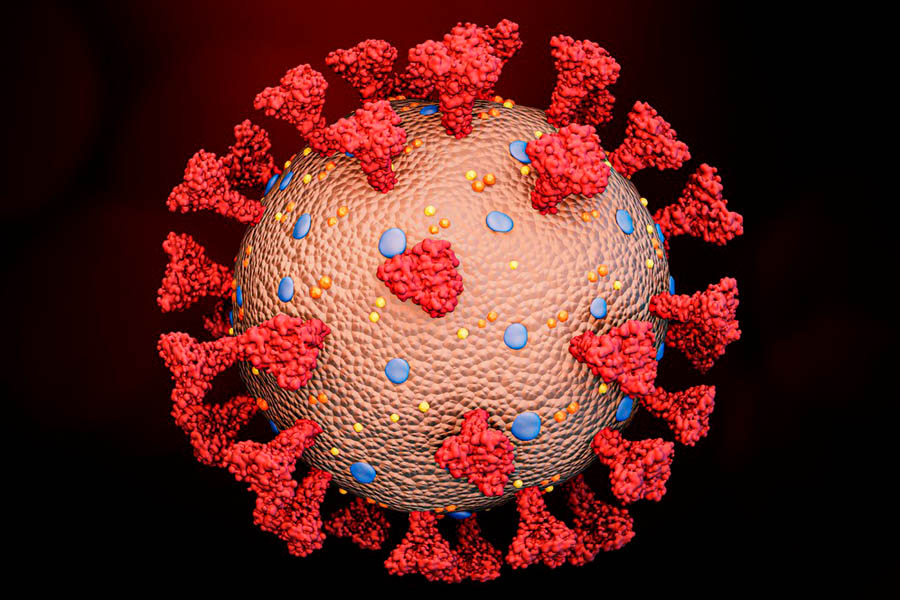
The SARS-CoV-2 virion consists of a nucleocapsid core enclosed by an envelope which contains three membrane proteins, namely spike (S), envelope (E) and membrane (M) [2]. The genome, with a size of around 30 kb, codifies for nonstructural replicase polyproteins and for the referred structural proteins [3]. The S protein is responsible for binding to the host cell-surface ACE2 receptor, initiating the infection [4,5]. After, the virus genome attaches to the host cell ribosomes, with the resulting translation of polyproteins, subsequently processed by proteolysis [2]. The virus lifecycle ends with the folding and packing of new virions, which infect other cells. E proteins are a group of small viral proteins that participate in the assembly and release of the virions [6] and are considered potential drug targets [2]. M proteins play a key role in the RNA packaging into the nucleocapsid [7], as well as N proteins [8].
The potential drug/vaccine targets
The S, E, M and N proteins, along with two isoforms of replicase polyprotein, namely 1a and 1ab, are considered potential drug/vaccine targets [2], and the S protein is already being used in several studies as a target antigen in vaccine development [9,10]. Nonetheless, the S protein undergoes conformational changes induced after its penetration in the host cell [11], and these changes seem to be provoked by mutations, which cause alterations in the antigenicity of this target. These dynamic changes in the protein may affect immune responses [12] and also may lead to drug resistance, if antiviral drugs are to be developed using S protein as target [13].
Hence, other proteins are being considered as targets for vaccine/drug development against SARS-CoV-2, and the N protein seems promising, as it is not affected by the variability of S protein. In fact, it was observed that N proteins from BAT-CoV, SARS-CoV and SARS-CoV-2 present highly conserved regions (N protein of SARS-CoV-2 shares nearly 90% sequence identity with that of SARS-CoV), so it is assumed that antibodies against the N protein of SARS-CoV would likely recognize the N protein of SARS-CoV-2 [2].
N-Protein specificities
N protein is a highly basic protein, containing two well-folded domains, both of them rich in β-strands. It presents highly positively charged regions which may facilitate binding to non-specific nucleic acids [14]. The function of N protein is to mediate viral assembly through interaction with the viral genome and M protein, helping to increase viral RNA transcription and replication [15]. Compared to the other virus proteins, N protein is more stable and fewer mutations occur over time [16].
IgG, IgA and IgM antibodies against the N protein were detected in confirmed COVID-19 patients’ sera [14,17] and were highly expressed during infection [15]. In addition, N protein was found to be very immunogenic, and the antibodies against N protein presented higher sensitivity and longer persistence than those against other structural proteins of SARS-CoV [18,19], eliciting proliferation and cytotoxic activity of SARS-specific T-cells [20,21].
Due to its multiple functions in viral lifecycle and infection, and the immunological response that it stimulates, N protein from SARS-CoV-2 is now a potential candidate as a target for a SARS-CoV-2 vaccine or drug.
Synthelis
Considering the potential of the N-protein from SARS-CoV-2 to fight COVID-19, Synthelis developed a protocol to produce this target in less than 2 weeks by using its cell-free protein expression system. After validation on rapid test devices from the market, this product is now available in Synthelis’ catalog.
As a service provider, we can also develop and produce almost any kind of proteins you may request for your research activity. Please contact us if you want additional information.
